Whether a Mastiff’s Cherry Eye Should Be Tucked
Few things are as instantly alarming to a Mastiff owner as seeing that angry, red bulge pop out of their gentle giant's eye. It appears suddenly, often without warning, turning your beloved dog's usually calm gaze into something that looks painful and utterly uncomfortable. This guide is here to walk you through exactly what "cherry eye" is, why the "tucking" procedure is almost always the best approach for your Mastiff, and what to expect when faced with this disconcerting eye condition.
Understanding Cherry Eye in Your Gentle Giant
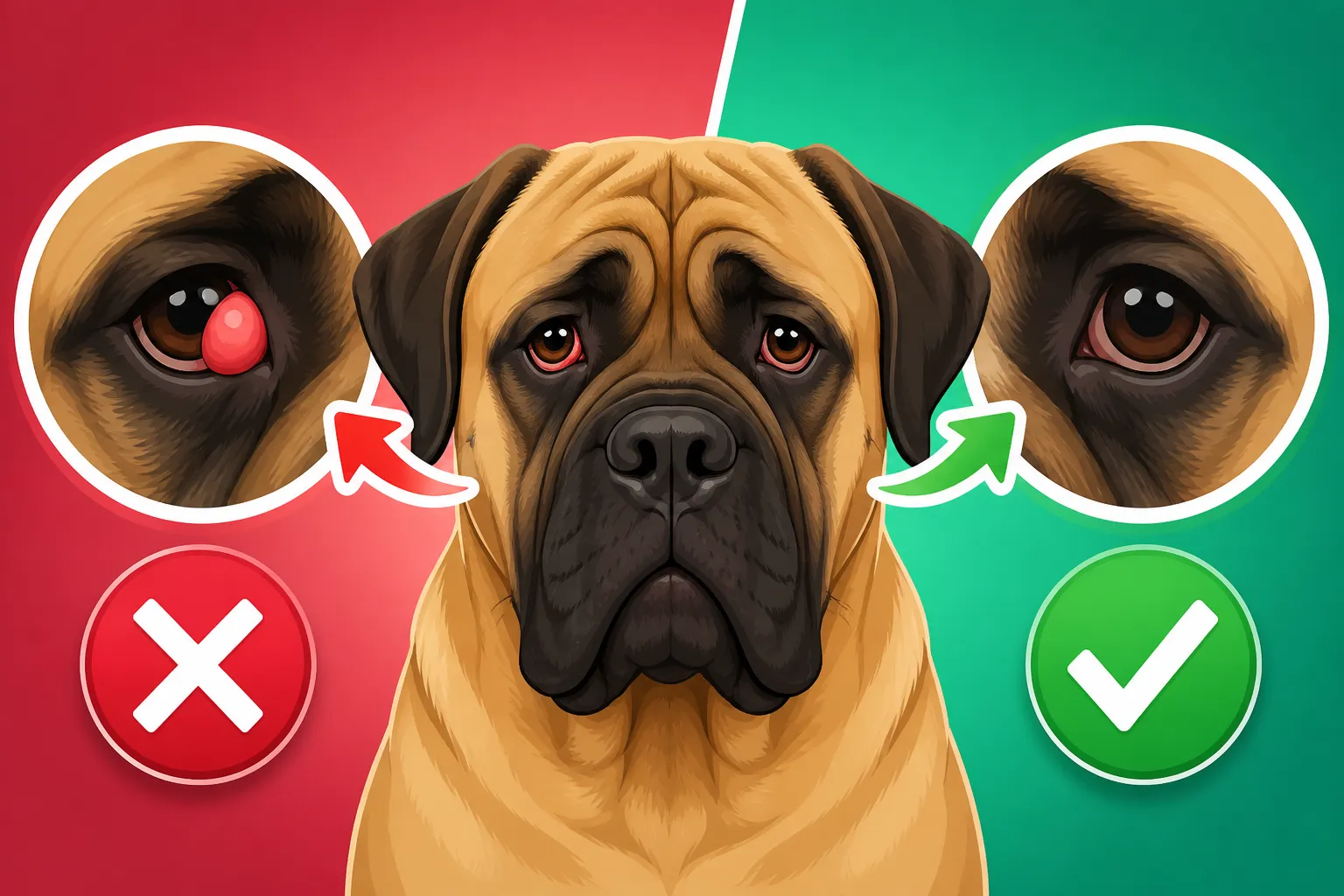
That unexpected red mass in the corner of your Mastiff's eye, often making them look perpetually surprised or even in pain, is what we commonly call "cherry eye." It’s not just a superficial irritation; it's a prolapse of the nictitating membrane gland, or the gland of the third eyelid. As an owner, your first instinct might be panic, followed by a strong desire to just push it back in – a natural reaction to seeing your dog uncomfortable.
What Exactly is Cherry Eye and Why Does it Happen in Mastiffs?
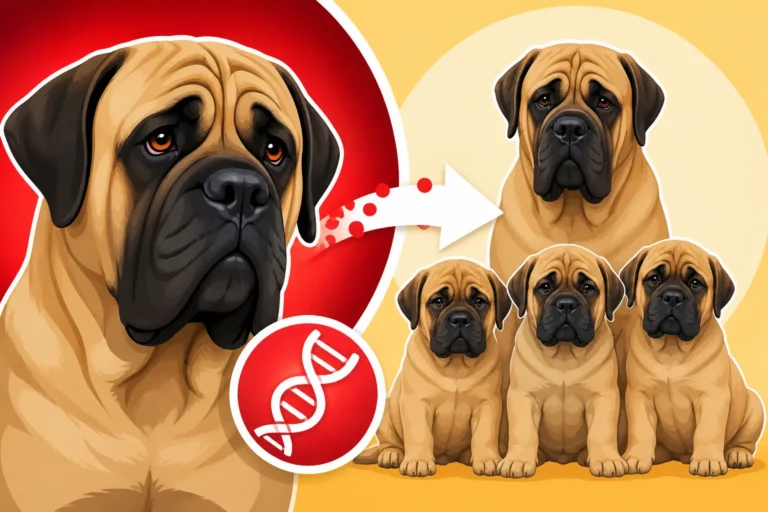
To understand cherry eye, let's talk about the third eyelid. Unlike humans, dogs have an extra eyelid, called the nictitating membrane, located in the inner corner of their eye. This membrane contains a tear-producing gland, crucial for keeping the eye lubricated and healthy. In a normal, healthy eye, this gland and membrane are neatly tucked away, barely visible. However, in certain breeds, including our magnificent Mastiffs, the connective tissue that holds this gland in place can be genetically weaker.
When this tissue fails, the gland can prolapse, meaning it pops out and becomes visible as a red, swollen mass. It's truly a sight that can make your heart sink. The exact cause isn't always clear, but genetics play a significant role. It's not usually caused by an injury, but rather an inherent predisposition. You might have seen it in puppies or young adult Mastiffs, but it can occur at any age. It’s an anatomical issue, not something you’ve done wrong, and it can happen to one eye or both. The size and shape of a Mastiff's head, with their sometimes looser skin and facial structure, might also contribute to this predisposition, making them more susceptible than some other breeds.
Common Symptoms and Initial Reactions
The most obvious symptom of cherry eye is, of course, the appearance of a distinct, round, reddish mass in the inner corner of your Mastiff's eye. It might look like a small, red cherry, hence the name. But beyond the visual, your Mastiff will likely show signs of discomfort. You might notice them pawing at their eye, blinking excessively, or rubbing their face against furniture or your leg. Their eye might also water more than usual, or you could see a thick, mucous-like discharge, especially if the exposed gland becomes irritated or infected.
Many owners experience a mix of alarm and frustration. "Is my dog in pain? What is that thing?" are common thoughts. It’s natural to feel a rush of concern for your Mastiff, who suddenly looks unwell and distressed. Some people try to gently push the gland back into place, and occasionally, it might even retract on its own for a short time. However, this is almost always temporary. The underlying structural weakness remains, and the gland will likely pop out again, often quite quickly. This is why addressing it properly is so important – it’s not just about aesthetics, it’s about preventing chronic irritation and protecting their vision and comfort long-term.
Surgical Options for Cherry Eye: Tucking vs. Excision
When faced with a Mastiff's cherry eye, the primary goal of any veterinary intervention is to relieve discomfort, restore normal eye function, and prevent future complications. There are two main approaches: repositioning (tucking) or, less ideally, removal (excision). For a breed like the Mastiff, which relies heavily on healthy tear production, the choice of procedure is critical.
The "Tucking" Procedure: Anchoring the Gland
The gold standard for treating cherry eye in Mastiffs, and indeed most breeds, is the "tucking" procedure, medically known as Nictitating Membrane Gland Repositioning (NMG-R). This technique is all about carefully repositioning the prolapsed gland back into its natural pocket behind the third eyelid and then anchoring it there with sutures. Think of it like a tiny, internal stitch that holds everything securely in place. The beauty of this method is that it preserves the gland, which is incredibly important for your Mastiff's long-term eye health.
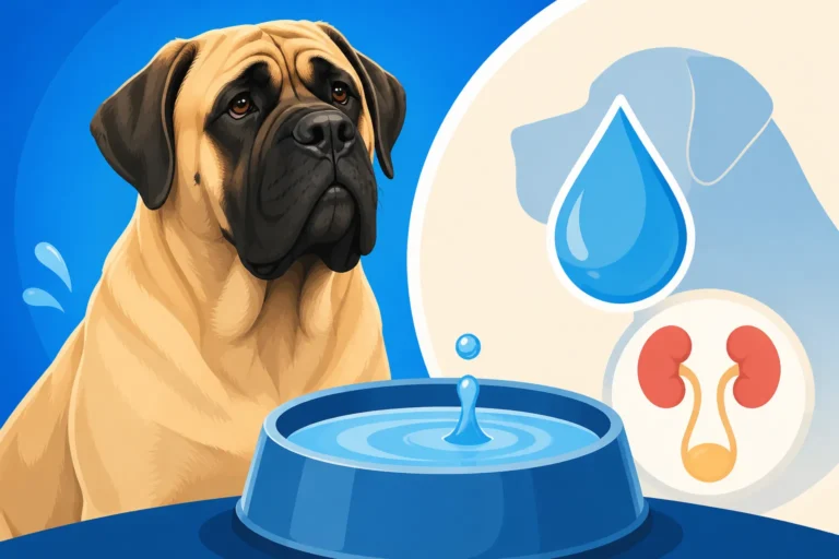
This gland is responsible for producing a significant portion of your Mastiff's tears – about 30-50% of the aqueous part of the tear film. Preserving this function is paramount, as removing it can lead to a condition called Keratoconjunctivitis Sicca (KCS), or "dry eye," which is a chronic, uncomfortable, and often expensive condition to manage for the rest of their lives. During the tucking surgery, your Mastiff will be under general anesthesia, which, for a massive breed, requires careful consideration and monitoring. You can learn more about Why Anesthesia Is Riskier in a Mastiff due to their size and physiological characteristics. The procedure itself is delicate but relatively quick, typically taking less than an hour per eye. After the gland is stitched into place, the sutures are usually absorbable and internal, meaning you won't see them.
Post-surgery, your Mastiff will need an Elizabethan collar (the "cone of shame") to prevent them from pawing at the sutures, which could lead to infection or undo the surgical repair. You'll also likely be sent home with eye drops or ointments to reduce inflammation and prevent infection, as well as oral pain medication. The recovery period usually involves careful monitoring and restricted activity for a couple of weeks. It’s a dedicated effort on your part, but knowing you’re preserving their tear function makes it worthwhile.
Gland Excision: A Last Resort and Its Risks
While "tucking" is the preferred method, it's worth understanding why gland excision – the complete removal of the prolapsed gland – is largely discouraged by veterinary ophthalmologists today. In years past, before the understanding of the gland's vital role in tear production became widespread, removal was a more common practice. However, we now know that taking out this essential tear gland has severe and often irreversible consequences for a dog's eye health.
The biggest risk of gland excision is the development of KCS, or chronic dry eye. Without the tear production from the nictitating membrane gland, the eye simply cannot produce enough tears to keep itself moist. This leads to chronic inflammation, irritation, corneal ulcers, and a thick, sticky discharge. Imagine living with perpetually dry, gritty eyes – it's incredibly uncomfortable. Managing KCS involves lifelong daily eye drops, typically a combination of tear stimulants and artificial tears, which can be time-consuming and costly. For a Mastiff, this means administering drops multiple times a day to a large, often unwilling patient. This chronic condition significantly impacts their quality of life and adds a considerable financial burden over their Six to Ten Years Is What a Mastiff Gets.
There are very, very few scenarios where excision might be considered, such as when the gland is so severely damaged or necrotic that it cannot be saved, or if multiple repositioning attempts have failed and the gland itself is no longer viable. Even in these rare cases, the veterinary team would discuss the high probability of KCS and the long-term commitment required to manage it. Always seek the advice of a board-certified veterinary ophthalmologist if gland excision is even suggested, as preserving the gland is almost always the better long-term solution for your Mastiff. The goal is always to avoid a future where your Mastiff needs constant medication just to keep their eyes comfortable and prevent vision loss.
The Pros and Cons of Tucking Cherry Eye
Deciding on surgery for your Mastiff is never a light matter. You want the best for them, and understanding the full scope of the procedure, including its advantages and potential drawbacks, is crucial. The "tucking" method for cherry eye, while highly recommended, comes with its own set of considerations.
Advantages of Surgical Repositioning
The primary and most significant advantage of surgically repositioning the prolapsed gland is the preservation of its tear-producing function. This cannot be stressed enough. Mastiffs, like all dogs, rely on healthy tear film to keep their eyes lubricated, clear away debris, and fight off infections. By keeping the gland intact, you dramatically reduce the risk of your Mastiff developing chronic dry eye (KCS) later in life. KCS is a miserable condition that requires daily, lifelong medication, which can become a huge burden for both you and your gentle giant. By tucking, you’re investing in your Mastiff's long-term comfort and ocular health.
When performed by an experienced veterinarian or, ideally, a veterinary ophthalmologist, the tucking procedure boasts a high success rate. Most Mastiffs recover well, and the eye returns to its normal, healthy appearance and function. The immediate discomfort your Mastiff felt from the exposed, irritated gland will be alleviated, improving their overall quality of life. Furthermore, a successfully tucked gland prevents the unsightly and potentially distracting "cherry" from being visible, restoring your Mastiff's majestic gaze. It also prevents the exposed gland from becoming chronically dry, inflamed, or infected, which can lead to more serious secondary issues like corneal ulcers.
Potential Drawbacks and Complications
While tucking is the preferred method, it's not entirely without its complexities. One of the most common concerns is the possibility of recurrence. Despite the best surgical technique, the gland can, unfortunately, prolapse again. This can happen if the sutures fail, if the underlying connective tissue remains too weak, or if the Mastiff is particularly prone to the condition. If one eye has cherry eye, there's also a significant chance that the other eye may develop it at some point, even if the first surgery was successful. This isn't a failure of the surgery on the first eye, but rather an indication of a bilateral genetic predisposition.
Another consideration, particularly for Mastiffs, is the general anesthetic. As giant breeds, Mastiffs require careful monitoring and specific anesthetic protocols. We touched on this with Why Anesthesia Is Riskier in a Mastiff, highlighting the need for a veterinary team experienced with large and giant breeds. Post-operative complications, though rare, can include swelling, bruising, infection at the surgical site, or the Mastiff managing to remove their protective cone and irritate the eye. It's crucial to follow all post-operative instructions meticulously to minimize these risks.
Finally, there's the financial aspect. Surgical correction of cherry eye, especially if performed by a specialist, can be a significant expense. This includes the initial consultation, pre-anesthetic bloodwork (which is always recommended for Whether a Mastiff Needs Pre Anesthetic Bloodwork), the surgery itself, medications, and follow-up appointments. This is where pet insurance, like a plan from Healthy Paws, can be an absolute lifesaver. While What Stone Surgery Costs on a Mastiff might differ, any major surgical intervention for a Mastiff can be costly, and being prepared or having a robust insurance policy can ease the burden. Understanding that The Mastiff's Short Life Buys the Biggest Bills means being financially ready for potential health issues from the start.
Making the Best Decision for Your Mastiff
Navigating a cherry eye diagnosis for your Mastiff can feel overwhelming, but with the right information and professional guidance, you can make the best decision for their health and comfort. Early intervention and informed choices are key to a successful outcome.
Consulting Your Veterinarian and Specialists
The moment you notice that tell-tale red bulge, your first step should always be to contact your primary veterinarian. They can confirm the diagnosis, assess the severity, and discuss initial management. However, given the delicate nature of eye surgery and the importance of preserving the tear gland, it's often highly beneficial to seek a referral to a board-certified veterinary ophthalmologist. These specialists have advanced training and experience specifically in eye conditions and surgeries, increasing the likelihood of a successful outcome, especially for a large breed like a Mastiff.
When consulting with your vet or a specialist, don't hesitate to ask questions. Inquire about their experience with cherry eye repairs in giant breeds, their preferred surgical technique (ensuring it's a tucking procedure that preserves the gland), and what success rates they typically see. Discuss the potential for recurrence and what the plan would be if it happened. They can also provide a detailed estimate of costs, which, as mentioned, can be substantial. Knowing these details upfront allows you to prepare adequately, both emotionally and financially. This might also be a good time to review your pet insurance coverage or consider obtaining a plan from providers like Healthy Paws, as unexpected surgeries can significantly impact your budget, echoing sentiments around What a Mastiff's Weight Does to Every Line on the Vet Invoice.
Preparing for Surgery and Post-Operative Care
Once you've decided on the tucking procedure, preparation is crucial for a smooth process. Your vet will likely recommend pre-surgical bloodwork to ensure your Mastiff is healthy enough for anesthesia, a vital step for such a large dog. On the day of surgery, follow all fasting instructions carefully. Make sure you have a quiet, comfortable space set up at home for your Mastiff's recovery, away from excessive activity or other pets. Consider a soft, wide Elizabethan collar (e-collar) rather than a rigid plastic one, which can be less comfortable for a giant breed.
Post-operative care is just as important as the surgery itself. Your Mastiff will come home with specific instructions regarding medications (eye drops/ointments, oral pain relief, possibly antibiotics) and activity restriction. It's paramount that they wear their e-collar continuously to prevent them from rubbing or scratching their eye, which could damage the surgical site. This can be challenging with a large dog, but their long-term eye health depends on it.
Watch for any signs of complications, such as excessive swelling, discharge, persistent pain, or if the gland appears to be prolapsing again. Promptly contact your veterinarian if you notice any concerns. Regular follow-up appointments will be scheduled to monitor healing and remove any non-absorbable sutures if used. Remember, while the initial recovery period is usually a couple of weeks, full healing and stabilization might take a bit longer. Investing in a successful surgery now can prevent a lifetime of chronic issues like Whether a Mastiff Gets Dry Eye, ensuring your Mastiff enjoys clear, comfortable vision for years to come.
Here's a quick comparison of the two primary approaches:
| Feature | Gland Repositioning ("Tucking") | Gland Excision (Removal) |
|---|---|---|
| Goal | Preserve tear-producing gland, restore normal anatomy. | Remove the gland completely. |
| Tear Production | Preserved (reduces dry eye risk). | Lost (high risk of KCS/dry eye). |
| Success Rate | High (with experienced surgeon). | Immediate resolution of "cherry" but high long-term complication rate. |
| Risk of Recurrence | Moderate (can happen in treated eye or other eye). | None for the treated eye, but KCS is a severe complication. |
| Complications | Anesthetic risk, post-op infection, suture reaction. | Anesthetic risk, lifelong chronic dry eye (KCS) requiring daily medication, corneal ulcers. |
| Long-Term Management | Minimal, usually none if successful. | Lifelong daily eye drops, frequent vet visits for KCS. |
| Cost Implications | Initial surgery cost, potential for second surgery if recurs. | Initial surgery cost, plus lifelong medication and vet visits for KCS. Pet insurance from providers like Healthy Paws is highly recommended for ongoing medical needs. |
| Recommended By | Veterinary ophthalmologists, general practitioners. | Rarely, only in extreme, irreparable gland damage cases. |
This table clearly illustrates why tucking is overwhelmingly the recommended approach for your Mastiff's cherry eye. It prioritizes their long-term health and comfort over a quicker, but ultimately more damaging, solution. For more on ocular health, consider reading about Eye and Eyelid Disease in a Mastiff.
Disclaimer
This article is intended for informational purposes only and does not constitute professional veterinary advice. Always consult with a qualified veterinarian or a board-certified veterinary ophthalmologist regarding any medical concerns or before making any decisions about your Mastiff's health and treatment. Individual outcomes may vary.